
Understanding the Complexity of the Knee Joint
The knee, a pivotal joint in the human body, is a marvel of biomechanical engineering. Composed of bones, ligaments, tendons, cartilage, and muscles, it facilitates a wide range of movements crucial for daily activities and athletic performance. This essay delves into the anatomy of the knee, exploring its intricate components and their roles in ensuring optimal functionality and health.
Bones: The Structural Foundation
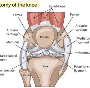
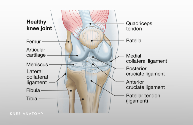
At the core of the knee joint lies a network of bones that form its structural foundation. The femur, tibia, and patella are the primary bones involved, each playing a distinct role in supporting weight, transmitting forces, and enabling movement. The femur, the thigh bone, articulates with the tibia, the shinbone, forming the main hinge of the knee. Meanwhile, the patella, or kneecap, protects the joint and enhances mechanical efficiency by providing leverage to the quadriceps tendon. Together, these bones create a stable yet flexible framework essential for mobility.
(For more detailed information on the bones of the knee, refer to this resource.)
Ligaments: Reinforcing Stability
Ligaments are fibrous bands of tissue that connect bones to each other, providing stability and limiting excessive movement within the joint. In the knee, four main ligaments—the anterior cruciate ligament (ACL), posterior cruciate ligament (PCL), medial collateral ligament (MCL), and lateral collateral ligament (LCL)—work in concert to maintain proper alignment and prevent dislocation.
- Anterior Cruciate Ligament (ACL): Situated within the knee joint, the ACL prevents the tibia from moving too far forward relative to the femur and controls rotational movements of the knee. It is particularly crucial for activities involving sudden stops, changes in direction, and pivoting motions, such as those common in sports like soccer, basketball, and skiing.
- Posterior Cruciate Ligament (PCL): Located within the knee joint, the PCL prevents the tibia from moving too far backward relative to the femur. It serves as a crucial stabilizer, especially during activities involving jumping, landing, and deceleration.
- Medial Collateral Ligament (MCL): Positioned on the inner side of the knee, the MCL provides stability by resisting forces that would push the knee inward (valgus stress). It is essential for maintaining proper alignment and preventing excessive medial joint opening.
- Lateral Collateral Ligament (LCL): Found on the outer side of the knee, the LCL reinforces stability by resisting forces that would push the knee outward (varus stress). It works in tandem with the MCL to maintain optimal joint alignment and prevent instability.
By reinforcing stability, these ligaments safeguard against injury and ensure smooth, controlled motion.
(To learn more about knee ligaments and their functions, visit this informative site.)
Tendons: Connecting Muscles to Bones
Tendons serve as the intermediary between muscles and bones, transmitting the forces generated by muscle contractions to produce movement. In the knee, several key tendons play vital roles in stabilizing the joint and facilitating ambulation. The quadriceps tendon connects the quadriceps muscles to the patella, enabling extension of the leg, while the patellar tendon attaches the patella to the tibia, facilitating movements such as jumping and kicking. Additionally, the hamstrings, located at the back of the thigh, are connected to the tibia via the pes anserinus tendon, contributing to knee flexion and stability. These tendons work synergistically to optimize biomechanical efficiency and promote joint integrity.
(For a deeper understanding of the role of tendons in knee function, refer to this resource.)
Cartilage: Cushioning and Smoothing Surfaces
Cartilage, a specialized connective tissue, lines the articulating surfaces of bones within the knee joint, serving as a shock absorber and reducing friction during movement. Two types of cartilage are found in the knee: articular cartilage and meniscal cartilage. Articular cartilage covers the ends of the femur, tibia, and patella, providing a smooth, low-friction surface that enables effortless gliding and weight distribution. Meanwhile, meniscal cartilage, located between the femur and tibia, enhances joint stability, absorbs impact, and aids in load-bearing. By cushioning and smoothing surfaces, cartilage preserves joint health and facilitates fluid motion.
(To explore the significance of cartilage in knee function, visit this informative site.)
Muscles: Powering Movement and Stability
Muscles surrounding the knee joint play a crucial role in powering movement, maintaining stability, and preventing injury. The quadriceps, located at the front of the thigh, extend the knee and stabilize it during activities such as walking, running, and jumping. Conversely, the hamstrings, situated at the back of the thigh, flex the knee and assist in hip extension, contributing to balance and agility. Additionally, the calf muscles, including the gastrocnemius and soleus, play a role in knee flexion and stability, particularly during activities involving weight-bearing and propulsion. By generating force and providing dynamic support, these muscles optimize knee function and enhance performance.
Understanding Knee Strength
Knee strength refers to the ability of the muscles surrounding the knee joint to generate force and provide dynamic support during movement. Strong muscles, including the quadriceps, hamstrings, calf muscles, and muscles of the hip and core, play a vital role in stabilizing the knee, absorbing impact, and controlling movement patterns. By enhancing muscle strength and endurance, individuals can mitigate excessive stress on the knee joint and reduce the risk of injuries such as ligament tears, tendonitis, and cartilage damage.
Effective Strategies for Knee Strengthening
1. Resistance Training
Incorporating resistance training exercises into your workout routine is an effective way to strengthen the muscles around the knee joint. Focus on exercises that target the quadriceps, hamstrings, and calf muscles, such as squats, lunges, leg presses, leg curls, and calf raises. Gradually increase the resistance or weight as your strength improves to continue challenging your muscles and promoting growth.
2. Functional Exercises
Include functional exercises that mimic real-life movements and activities to improve knee stability and functional strength. Examples include step-ups, lateral lunges, single-leg balance exercises, and agility drills. These exercises help improve balance, coordination, and proprioception, enhancing joint stability and reducing the risk of falls and injuries.
3. Flexibility and Mobility Work
Incorporate stretching and mobility exercises into your routine to maintain flexibility and range of motion in the knee joint and surrounding muscles. Focus on stretching the quadriceps, hamstrings, calves, hip flexors, and iliotibial (IT) band to prevent muscle imbalances and reduce the risk of overuse injuries. Foam rolling and self-myofascial release techniques can also help alleviate muscle tension and improve tissue quality.
4. Balance and Stability Training
Include balance and stability exercises to challenge the muscles that support the knee joint and improve proprioception. Utilize balance boards, stability balls, Bosu balls, and proprioceptive pads to perform exercises such as single-leg stands, stability lunges, and stability ball squats. These exercises help activate the stabilizing muscles around the knee and improve neuromuscular control, reducing the risk of instability-related injuries.
5. Core Strengthening
Don’t neglect the importance of core strength in supporting proper alignment and stability of the knee joint. Incorporate exercises that target the core muscles, including the abdominals, obliques, and lower back, such as planks, Russian twists, bicycle crunches, and stability ball rollouts. A strong core helps maintain proper posture, distribute forces more evenly throughout the body, and reduce the risk of compensatory movements that can lead to knee injuries.
6. Gradual Progression and Proper Form
When engaging in knee strengthening exercises, prioritize proper form and technique to maximize effectiveness and minimize the risk of injury. Start with light resistance and gradually increase intensity and load as your strength improves. Avoid rapid progressions or overexertion, as this can increase the risk of muscle strain or joint injury. Listen to your body and modify exercises as needed to avoid pain or discomfort.
The Knee: Common Ligament Injuries and Their Impact
The knee joint, a complex structure comprising bones, ligaments, tendons, cartilage, and muscles, is prone to various injuries due to its pivotal role in weight-bearing and mobility. Among the most prevalent knee injuries are ligament tears, which can significantly impair function and lead to long-term complications. This essay explores common ligament injuries of the knee, their causes, symptoms, and implications for individuals affected by them.
Understanding Ligament Injuries
Ligaments are tough bands of fibrous tissue that connect bones to each other, providing stability and limiting excessive movement within the joint. When subjected to sudden or excessive forces, such as those encountered during sports activities or traumatic accidents, ligaments can tear or become stretched beyond their normal limits, resulting in injury. The four primary ligaments of the knee—the anterior cruciate ligament (ACL), posterior cruciate ligament (PCL), medial collateral ligament (MCL), and lateral collateral ligament (LCL)—are particularly susceptible to damage.
Anterior Cruciate Ligament (ACL) Injury
The ACL is one of the most commonly injured ligaments in the knee, often occurring during activities involving sudden stops, changes in direction, or pivoting motions. ACL tears typically result from non-contact mechanisms, such as landing awkwardly from a jump or abruptly changing direction while running. Symptoms of an ACL injury may include a popping sensation at the time of injury, swelling, instability, and difficulty bearing weight on the affected knee.
Posterior Cruciate Ligament (PCL) Injury
PCL injuries are less common than ACL injuries but can still occur, particularly in high-impact sports or motor vehicle accidents where the knee is forcefully bent or hyperextended. Unlike ACL injuries, which often result from non-contact mechanisms, PCL tears typically occur due to direct impact to the front of the knee while it is bent. Symptoms of a PCL injury may include swelling, pain at the back of the knee, instability, and difficulty walking or bending the knee.
Medial Collateral Ligament (MCL) Injury
The MCL is located on the inner side of the knee and is frequently injured due to direct blows to the outer side of the knee or forces that push the knee inward (valgus stress). MCL injuries are common in contact sports, such as football or rugby, where players may sustain lateral blows to the knee during tackles or collisions. Symptoms of an MCL injury may include pain and tenderness on the inner side of the knee, swelling, instability, and difficulty bending or straightening the knee.
Lateral Collateral Ligament (LCL) Injury
The LCL is situated on the outer side of the knee and is less frequently injured than the MCL due to its inherent strength and stability. LCL injuries typically occur as a result of direct blows to the inner side of the knee or forces that push the knee outward (varus stress). While less common than injuries to other knee ligaments, LCL tears can still occur, particularly in sports or activities involving sudden changes in direction or collisions. Symptoms of an LCL injury may include pain and tenderness on the outer side of the knee, swelling, instability, and difficulty bearing weight on the affected leg.
Implications and Treatment
Ligament injuries of the knee can have significant implications for affected individuals, ranging from pain and swelling to instability and functional limitations. Prompt diagnosis and appropriate management are crucial for optimizing outcomes and preventing long-term complications. Treatment for ligament injuries may vary depending on the severity and nature of the injury but often includes a combination of conservative measures such as rest, ice, compression, and elevation (RICE), physical therapy, bracing, and in some cases, surgical intervention.
Conservative Management
For mild to moderate ligament injuries, conservative management may be sufficient to promote healing and restore function. This may involve a period of rest followed by gradual rehabilitation exercises aimed at improving strength, flexibility, and proprioception. Physical therapy plays a key role in rehabilitating the injured knee, helping individuals regain stability, range of motion, and functional mobility. Additionally, the use of supportive braces or orthotics may be recommended to provide external stabilization and protect the injured ligament during activities.
Surgical Intervention
In cases of severe ligament injuries or failure to respond to conservative treatment, surgical intervention may be necessary to repair or reconstruct the damaged ligament. Surgical techniques for ligament reconstruction typically involve using grafts, often from the patient’s own tissue (autograft) or from a donor (allograft), to replace the torn ligament and restore stability to the knee joint. Rehabilitation following surgery is essential for optimizing outcomes and may involve a structured program supervised by a physical therapist to gradually reintroduce weight-bearing activities and restore strength and function
Knee joint
The knee joint is a masterpiece of biomechanical complexity, comprising bones, ligaments, tendons, cartilage, and muscles that work harmoniously to facilitate movement and ensure stability. Understanding the anatomy of the knee is paramount for maintaining its health and preventing injury, as it empowers individuals to make informed decisions regarding exercise, rehabilitation, and lifestyle choices. By appreciating the vital components that comprise this remarkable joint, we can strive to optimize its functionality and longevity, enabling us to pursue our passions and live life to the fullest.